Introduction
Prostate cancer may be a concern if the patient’s PSA test is abnormally raised. If the urologist suspects that the patient may have cancer, he may recommend that the patient go for prostate biopsy. The standard prostate biopsy involves placing an ultrasound probe into the rectum, and tiny samples of prostate tissue are obtained using a biopsy needle. Usually 12 or more different areas of the prostate are sampled in a systematic manner.
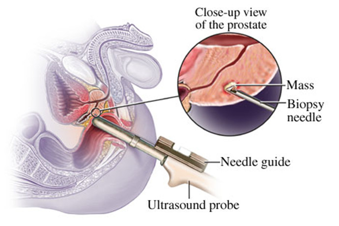
Prostate biopsy is performed through the rectum
Most of the time, the ultrasound does not show where the cancer is, so the urologist has to take enough samples to ensure that most of the prostate are sampled. An interesting fact is that prostate cancer is the only cancer diagnosed by biopsy without the tumour actually being seen.
There are some problems with the current biopsy technique:
- It is possible that the cancerous area is missed during the biopsy. Sometimes a patient has to go for repeat biopsy because the initial biopsy does not show cancer but his PSA continues to increase.
- Obtaining biopsies from multiple sites may increase the chance of diagnosis of clinically insignificant prostate cancer, ie cancer which may not have caused harm to the patient in his lifetime. This may lead to unnecessary anxiety and potentially unnecessary treatment.
MR/ultrasound fusion guided biopsy of the prostate
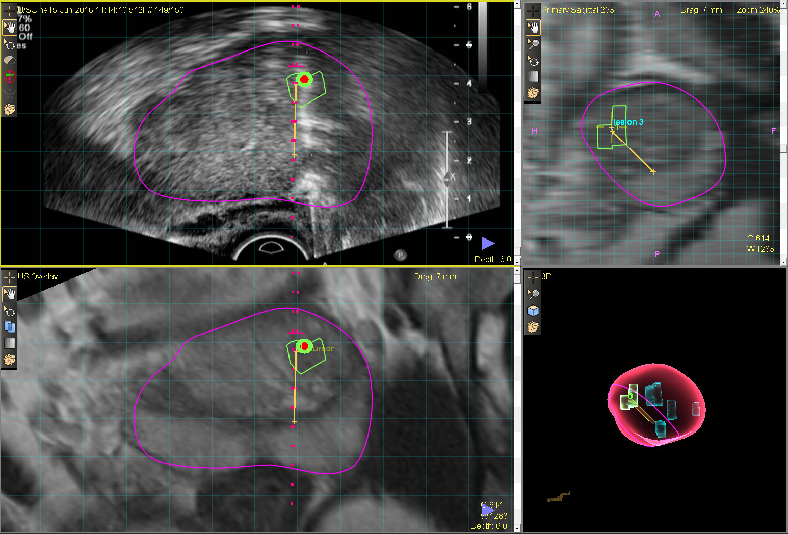
Using MRI scans, doctors are now better able to tell which areas of the prostate are more likely to have cancer, and studies have shown that suspicious areas on MRI are more likely to harbor significant cancer. With the introduction of MR/ultrasound fusion guided biopsy of the prostate, it is now possible to accurately biopsy lesions in the prostate that are shown on MRI. The lesions in the prostate are first outlined on the MRI images. Next, transrectal ultrasound is performed. Fusion of the MR and ultrasound images takes place and the outlined lesions are superimposed live over the ultrasound images. The targeted lesions are then biopsied. In most instances, additional biopsies are also obtained from normal-looking areas to make the sampling more complete.

MRI-US fusion guided biopsy platform

Targeted biopsy of a suspicious area of the prostate
Advantages of MR/ultrasound fusion guided biopsy of the prostate
- Targeted biopsy increases detection of high risk cancers and reduces detection of clinically insignificant cancers. This is better as men with high risk cancers are the ones who would benefit the most from treatment.
- It improves detection of cancers in men who had previous negative prostate biopsy. This may help to reduce the number of additional biopsy sessions that these men may otherwise be subjected to.
- It facilitates repeat biopsy in patients on active surveillance for prostate cancer; as it allows repeat biopsy of areas of the prostate where cancer was previously found.
The downside
The downside is the cost involved, due to the need for MRI and the specialized equipment involved in the biopsy. The cost of MR/ultrasound fusion guided biopsy of the prostate may be balanced out by the savings from not having to undergo repeat biopsy. Costs may also come down as the technique becomes more widespread and equipment becomes less expensive.
Conclusion
MR/ultrasound fusion guided biopsy of the prostate allows the urologist to biopsy the prostate more accurately, and is a worthwhile improvement over the conventional method of systematic biopsy.